top of page

Our Expert

Nina Fedosova

Master of Mathematics, Leningrad State University

Academy of Methods and Technologies of Management (LIMTU)

With over 30 years of experience in the IT industry, certified IT architect and technical expert (TOGAF, IBM, ITIL) with a proven track record in delivering complex IT projects. Brought deep expertise in LAN and SAN technologies, operating systems across multiple server platforms, and high-availability cluster solutions. Career spans the full project lifecycle—from design to implementation and support—ensuring practical, reliable, and scalable outcomes. A strong and dedicated technical leader, continuously seek and solve new challenges in IT and AI.

Nina.jpeg

Our Product

Developed AI-powered software prototype assists pathologists in the differential diagnosis of oncological diseases.

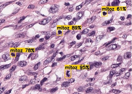
It leverages deep learning (ResNet architecture with residual blocks) to automate the detection of pathological mitoses and other key features on digitized histological slides.

Also it enables high-throughput, automated image analysis, reducing workload, increasing diagnostic accuracy, and minimizing human errors.

Current model characteristics (based on the functional assessment, clinical trial is still to be done):

  • Precision = 96%

  • Recall = 98%

  • Accuracy = 97%

MegAITex

Призначення лікаря

Accurate diagnosis on cancer is one of the most complex areas in clinical oncology due to the rarity, diversity, and heterogeneity of these tumours.

Current diagnostic processes are time-consuming, require significant manual labour, and are subject to expert disagreement, especially in differentiating benign from low-grade malignant tumours.

There is a lack of industrial solutions for automated recognition of morphological and histological images using AI, and no high-quality, open-access datasets for histological image analysis.

Current accuracy is 97% with potential for growth.

What We Do

Picture2.jpg
Picture1.jpg
Picture3.jpg
Picture4.jpg

*Atypical mitosis is a disruption of the normal process of cell division (called mitosis), which leads to abnormal distribution of genetic material between the two new cells. As a result, the daughter cells may receive an unequal number of chromosomes or have chromosomal abnormalities.

Innovative AI model development approach for the atypical mitoses* detections.


A fundamental distinction of our solution are graphics processing technologies and specially developed mathematical methods for object detection with Medical science and experts' engagement.


Our technology uses advanced ML architecture improved and trained as a new model which enhanced and customized for other fields of medicine diagnostics.


Developed mathematical model was tested in State Medical Institution and further fine-tuned to minimize
the number of false positives. For this purpose, individual categories of images that appeared “similar” from the models' perspective were separated into subcategories.

Neural Network Architecture

Technology and Innovation

Neural Network Architecture

Utilizes ResNet-50 and Feature Pyramid Network (FPN) for robust feature extraction and small object detection on large-scale images.

Handles massive histological images (up to 1.5 GB), segments them into manageable tiles for efficient analysis.

Data Processing

Novelty

Focuses on object detection (not just classification or segmentation), overcoming limitations of existing AI models in clinical practice.

Our Consumers

Рецепт

Medical institutions
specializing in diagnostics

Лабораторна робота

Manufacturers
of medical equipment
and analytical systems

Вчений біля мікроскопа

Research medical institutions

Our Mission

Revolutionize cancer diagnostics by harnessing the power of advanced AI and medical science to deliver faster, more accurate, and reliable pathology insights.

We are committed to empowering clinicians with cutting-edge tools that reduce diagnostic errors, enhance efficiency, and ultimately improve patient outcomes. By combining innovation in artificial intelligence with deep medical expertise, we aim to close the gap in histopathological analysis and set a new global standard for accessible, scalable, and precise medical diagnostics

Early and accurate diagnostics
improves the patients' quality of life

Любіть Світ
Руки в рукавичках тримають флакони
Samples
Автоматизоване виробництво вакцин
В ЛАБОРАТУРІ

Our Adviser

GN1.jpeg

Dr. Gennady Nikolaevich Berchenko, MD, Professor

Head of the Department of Pathological Anatomy at the N.N. Priorov Central Institute of Traumatology and Orthopedics (CITO), where he has worked since 1988. A Doctor of Medical Sciences and Professor of Pathological Anatomy, he holds the highest qualifications in both pathological anatomy and clinical laboratory diagnostics.

With over 30 years of experience, he is a leading expert in bone and joint pathology, regenerative processes, and the use of novel biomaterials in traumatology and orthopedics.

Dr. Berchenko is the author of more than 500 scientific publications and has supervised 23 PhD dissertations.

Our Publications

  • ABSTRACT

    BACKGROUND: Modern computer systems allow digitizing and examining images of histological preparations, which led the authors to the idea of using machine learning tools in digital pathohistology. The ability of neural networks to find sub-visual image features in digitized histological preparations provides the basis for better qualitative and quantitative image analysis. Existing machine learning methods provide good accuracy and speed in recognizing various images, which gives hope for their wide application, including in oncologic diagnostics.

    AIM: Use methods of mathematical modeling to identify pathological mitoses in histological preparations as the main sign of the difference between malignant and benign tumor growth.

    MATERIALS AND METHODS: Histological images of the N.N. Priorov National Medical Research Center of Traumatology and Orthopedics were used as a data set for the neural network model. The model was tested using 188 histologic slides from 67 patients treated at the institute. Histological preparations were scanned on a Leica Aperio CS2 microscope with a ×400 resolution and converted into JPEG format with further processing. Next, the test images were analyzed in streaming mode using the created neural network model in order to obtain the coordinates of the desired diagnostic object — pathological mitosis and the probability with which the model found the object of this category. The obtained images were analyzed by a pathologist to determine whether the detected object corresponded to pathological mitosis.

    RESULTS: The authors have chosen an architecture, developed a methodology for training a neural network, and created a model that can be used to detect pathologic mitoses in histologic preparations. The authors do not attempt to replace the physician, but show the possibility of an integrated approach to data analysis by a computer system and a pathologist.

    CONCLUSIONS: The developed mathematical model of neural network used as part of technological solution for recognizing pathological mitoses in scanned histological preparations can be used as a tool to reduce the time of research and increase the accuracy of diagnosis by a pathologist.

    Keywords: neural network; mathematical model; artificial intelligence; tumor; pathological mitosis; machine learning; bone pathology.

    Keywords: neural network; mathematical model; artificial intelligence; tumor; pathological mitosis; machine learning; bone pathology.

    Berchenko, G. N., Fedosova, N. V., Kochan, M. G., & Mashoshin, D. V. (2024). Neural network model development for detecting atypical mitoses in histological slides. N.N. Priorov Journal of Traumatology and Orthopedics, 31(3), 337–349.

  • ABSTRACT
    The problem of improving the quality of a convolutional neural network (CNN) in the case of searching for objects on histological scans has been around for a long time and comes down, first of all, to choosing the correct CNN scheme and preparing a dataset of stable quality. The operation of the object detection algorithm is influenced by many factors, including image quality, image size, and the search for the object itself. Search for modern studies demonstrating the influence of various image characteristics on the selection of training programs and the choice of CNN design on the quality of the created model. As research basis, the literature for the past 5 years devoted to data preprocessing, methodologies, requirements for images included in datasets, creating images for CNN models, and structure selection issues were analyzed. At the time of the study, the requirements for image sizes were not formulated and there is no data on the sizes of objects to the image sizes for optimizing the model. In addition, the problem of choosing a neural network scheme is not transparent and algorithmic. In most cases, researchers use the structures they developed or used themselves, without explaining the reasons or criteria for their choice, or comparing alternatives. All these issues complicate the process of developing CNN models for digital image processing. Current research presented and provided a brief overview of studies on preparing images for a dataset and possible choice of CNN structure.

    Keywords: neural network; mathematical model; artificial intelligence; machine learning.

    Fedosova, N. V., Berchenko, G. N., Shugaeva, O. B., Mashoshin, D. V., & Kochan, M. G. (2024). The influence of CNN architecture, image size and quality to object detection model on histological specimens. N.N. Priorov Journal of Traumatology and Orthopedics, 31(4).

  • Application of neural network for morphological assessment of revascularized autograft remodeling at the stage of implant placement in jaw defects

    Abstract

    Relevance. The study examines the possibility of using a mathematic model of an artificial neural network (ANN) for comparative quantitative morphological assessment of revascularized autograft remodeling after reconstructive surgery in patients with jaw defects.

    Materials and methods. Jawbone biopsies of 30 patients were histologically examined during the implant placement stage, 6, 9, 12 months after the reconstructive surgery with a revascularized fibular graft. Histology images were analyzed using a mathematic model created on the basis of GoogLeNet and trained on morphological images of bone maturation. Histology slides were digitized by a scanning microscope Leica SC2 and streamed through the neural network model.

    Results. During the follow-up period, jaw defect areas demonstrated relatively mature bone tissue formation with various intensity rates of remodeling and maturation of the newly-formed bone. At that, the results of the descriptive histology were consistent with the quantitative results of the ANN mathematic model, created on the basis of the software-hardware system developed by the authors. The confirmed significance level is 95% or higher.

    Conclusion. Pathomorphological data of biopsies were studied using an ANN mathematical model, built on a software-hardware system, which allowed analyzing all microscope fields of view of a histological slide, bypassing random samples, as well as the evaluation by a pathologist of individual microscope fields of view, to exclude the possibility of unrepresentative sampling and the influence of human factor, which significantly increases the significance of the received results.

    Key words: vascularized fibular autograft, dental implantation, morphological study, autograft remodeling, mathematic model, artificial neural network

    Berchenko, G. N., Braylovskya, T. V., Fedosova, N. V., & Tangieva, Z. A. (2021). Application of neural network for morphological assessment of revascularized autograft remodeling at the stage of implant placement in jaw defects. Parodontologiya, 26(3), 188–196.

  • Development of the mathematical model neural network for morphological assessment of repair and remodeling of bone defect

    Currently, there is no longer any doubt that the use of artificial intelligence models has exceptional potential in many areas of our life, including medicine. It brings medical research to a fundamentally new qualitative level due to a high degree of accuracy in the analysis of growing volumes of medical data, avoiding the influence of the human factor and related medical mistakes. Despite the rapid development of neural networks, their practical application in modern scientific research is extremely rare. In the articles of scientists, there are no works in which neural networks used for analytics of morphological images.

    The methods of mathematical statistics currently used for this purpose are very complex and, in most cases, difficult for medical scientists to apply. This leads to many errors and, in some cases, to unscientific and absurd conclusions. Therefore, the authors of this work have developed methodology of creation the mathematical model based on GoogLeNet architecture, which used for morphological healing process of a bone defect investigation.

    The expert pathologist confirms results of morphological investigation conducted by mathematical model created based on a convolutional artificial neural network. The reliability of the results of a qualitative and quantitative morphological study - image analysis using the neural network developed by the authors of the article - significantly exceeds the reliability of the processing of the results by a specialist performed in the traditional way.

    The mathematical model makes it possible to exclude the random sampling, as well as the human factor in evaluating research results.

    Key words: neural network GoogLeNet, artificial intelligence, mathematical model, bone defect healing

    Fedosova, N. V., Berchenko, G. N., & Mashoshin, D. V. (2021). Development of the mathematical model neural network for morphological assessment of repair and remodeling of bone defect. Mathematical Modeling, 33(9), 22–34.

Want to know more about our product?

bottom of page